You may be familiar with the abdominal ultrasound scan, where the sonography is done over your abdomen.
In a transvaginal (internal) ultrasound scan, the scanning probe is placed inside the vagina and is closer to the organs being examined.
This type of scan helps provide clearer pictures of the uterus, ovaries and surrounding structures in the lower abdomen than Abdominal USG.
Why and When
- Please note that an internal scan can be performed at any time during a woman’s life – in pregnancy, during your period or after menopause.
- You will not be asked to have a vaginal ultrasound scan if you are unable to tolerate vaginal (internal) examinations or if you are not sexually active and are a virgin.
- We use this method of scanning to help assess the cause of problems such as infertility or abnormal vaginal bleeding when we need to make a better assessment and more accurate measurement of the lining of the womb (endometrium).
- We also perform this type of scan if you have been experiencing a problem like pelvic pain and we need to make a better assessment of the ovaries and tubes to look for cysts or any other conditions that may be affecting these organs.
- It is routinely suggested as the method of assessment of Early Pregnancy below 10 weeks gestation in a much better and clear way, particularly for assessing pregnancy complications like miscarriage or ectopic pregnancy.
How is the scan performed?
- You do not need a full bladder for this scan so you will need to empty your bladder completely prior to the scan.
- Once you have emptied your bladder, we will ask you to undress completely from the waist down and put on a cover over your waist with a sheet which we will give you. If you are wearing a skirt or sari, you may prefer to just remove your underwear. If you are using a tampon or a vulval pad, this will need to be removed before the scan.
- You will be asked to lie on your back on the ultrasound couch and position yourself in a way that allows the scan to be performed easily. This will involve raising your knees and keeping the soles of your feet flat on the ultrasound couch. A specially designed ultrasound probe is used for this procedure. It will be covered with a protective sheath and lubricating gel, then gently inserted into your vagina. The ultrasound probe will need to be moved in different positions in order to visualize the uterus and ovaries clearly.
- Your scan appointment is usually booked for 15 minutes but this may vary depending on how clear the pictures are or if we need to do other investigations.
- In most cases, the report will be given within 15 minutes after the procedure.
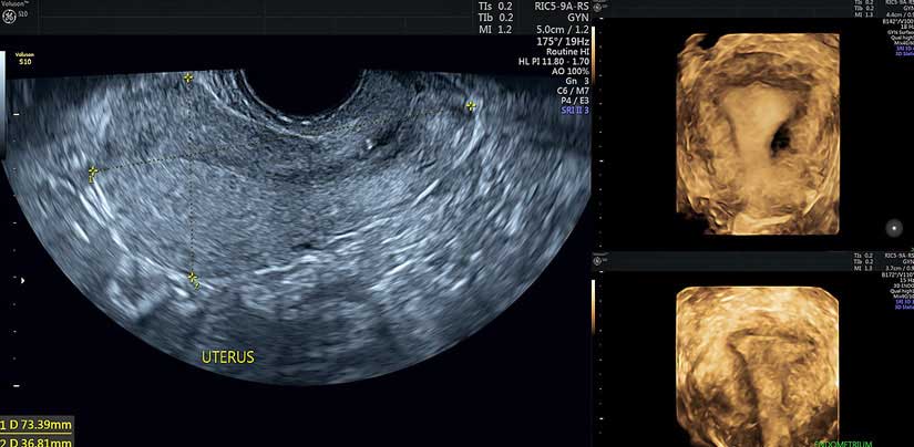
Why 3D Ultrasound
- Three-dimensional ultrasound (3D USG) is a fast-evolving imaging technique that holds great potential for use in gynaecology. Patients can benefit from adding 3D USG to their routine gynaecological workup as it provides fast and accurate results in a relatively cost-effective way, compared to CT Scan or MRI.
- As in two-dimensional ultrasound (2D USG), the transvaginal approach is preferred in gynaecologic examination with 3D USG.
We use 3D technology regularly when appropriate in our transvaginal USG examination at no additional cost.
Here are some of the reasons for specially using the 3 D USG by TVS
- Detection of congenital uterine anomalies
- Defining and mapping uterine lesions such as fibroids, very valuable when making clinical decisions and surgical planning.
- Assessing the Ovaries by Sonography-based Automated
Volume Calculation (Sono AVC) is one for counting antral follicles and monitoring follicular growth
If you have any concerns about this procedure, please discuss this with the Doctor who will be performing the examination, prior to the procedure.

Download PDF