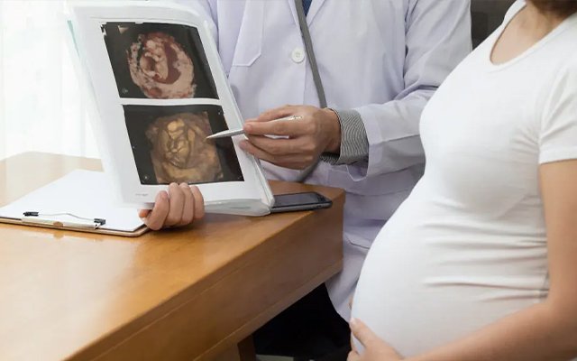
Wondering if your baby is growing just right? Every parent wants reassurance that their little one is developing healthily—and that’s where pregnancy ultrasound plays a pivotal role. This isn’t just a routine check; it’s a window into your baby’s world, revealing heartbeats, tiny limbs, and the formation of vital organs.
From the moment a heartbeat is detected to detailed scans of the spine, brain, and kidneys, ultrasound imaging gives doctors and parents a real-time view of growth patterns and overall well-being. It transforms the invisible into a vivid story of development, helping spot potential concerns early and ensuring timely guidance throughout pregnancy.
In this guide, we’ll explore how doctors track your baby’s growth, the methods used, and what each scan tells you. We’ll break it all down into simple, relatable insights while showing why regular monitoring is essential for a healthy pregnancy journey.
What Is a Pregnancy Ultrasound?
A pregnancy ultrasound uses high‑frequency sound waves to create detailed images of your baby inside the womb. Unlike X‑rays or other imaging technologies, ultrasounds are considered safe when performed by trained professionals because they don’t use ionising radiation. Ultrasounds form the backbone of antenatal care and are used throughout pregnancy to assess structure, size, and growth patterns.
Doctors schedule routine ultrasounds at key gestational ages to track development and detect potential concerns. These scans provide objective data on fetal health and give expectant parents a first glimpse of their baby’s growth.
At Fetomat Wellness, pregnancy ultrasound in Kolkata is more than just a scan. It is a careful assessment of your baby’s growth, development, position, heartbeat, and overall well-being, guided by experienced fetal medicine specialists.
Why Ultrasound Is Essential in Prenatal Care

Ultrasound plays a critical role in:
- Confirming pregnancy and fetal heartbeat
- Estimating gestational age and due date
- Monitoring the anatomical development of organs and structures
- Assessing fetal growth trends over time
- Detecting anomalies early
- Evaluating placenta and amniotic fluid levels
- Planning management for high‑risk pregnancies
Without ultrasound imaging, many of these insights simply wouldn’t be possible with the same accuracy or safety.
Key Methods Doctors Use to Detect Baby’s Growth
Doctors use several evidence‑based ultrasound techniques that allow precise assessment of fetal development. Let’s break them down:
1. Fetal Biometry: The Core Growth Check
Fetal biometry involves measuring key anatomical parameters that reflect growth patterns:
| Measurement | What It Represents |
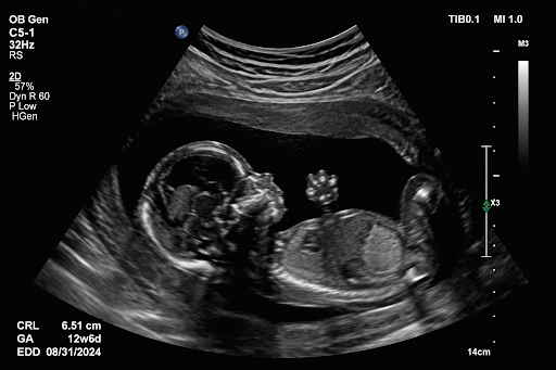
| CRL (Crown‑Rump Length) | Early pregnancy size estimate |
| BPD (Biparietal Diameter) | Head width |
| HC (Head Circumference) | Overall head size |
| AC (Abdominal Circumference) | Belly size |
| FL (Femur Length) | Length of thigh bone |
These measurements help estimate gestational age and assess whether fetal size is appropriate for that age. If measurements vary significantly from expected norms, further monitoring or tests may be recommended.
During a scan, a technician captures these measurements and compares them to established growth charts based on thousands of healthy pregnancies. Trends over time are often more informative than single measurements.
2. Anatomy Scans: Beyond Size — Structural Assessment
Around 18–22 weeks of pregnancy, a more detailed “anatomy scan” is performed. This scan checks major organs and structures, such as the brain, heart, spine, kidneys, and limbs. It detects structural anomalies that may not be apparent in early scans.
This comprehensive assessment supports a full view of growth and development—not just size.
3. Doppler Ultrasound: Checking Blood Flow and Heart Health
Doppler ultrasound tools assess blood flow within the umbilical cord and certain fetal vessels. This helps determine whether your baby is receiving enough oxygenated blood—a key indicator of well‑being. Abnormal blood flow patterns may indicate growth restriction or placental issues.
4. Serial Scans: Monitoring Growth Over Time
A single ultrasound gives a snapshot—but serial scans tell a story. By comparing multiple scans over weeks, doctors can see growth trajectories and identify concerns like intrauterine growth restriction (IUGR), allowing timely intervention.
What Each Trimester’s Ultrasound Tells You
First Trimester (up to ~13 weeks)
- Confirms viability
- Detects fetal heartbeat
- Measures CRL to estimate gestational age
- Determines number of fetuses
Mid‑Pregnancy (18–22 weeks)
- Detailed anatomy scan
- Checks organ development
- Assesses spine, brain, heart structure
Third Trimester
- Monitors ongoing growth
- Checks placenta and amniotic fluid
- Detects signs of growth abnormalities
Ultrasound measurements aren’t just numbers—they give doctors a precise language to understand and forecast your baby’s development.
When Additional Testing Might Be Needed
Ultrasound may indicate the need for further testing when:
- Fetal measurements don’t match expected gestational age
- Blood flow patterns raise concerns
- The placenta is positioned unusually
- Amniotic fluid levels are abnormal
In these cases, doctors may use Doppler studies, repeat scans, or other diagnostic procedures to gather more information.
How Ultrasound Guides Pregnancy Management and Outcomes
Ultrasound provides clinical data that can significantly influence decisions around:
- Timing of delivery
- Need for maternal or fetal interventions
- Monitoring high‑risk pregnancies
- Detecting complications early
This helps healthcare teams personalise care plans unique to each pregnancy. Connect with Fetomat Wellness for trusted pregnancy ultrasound in Kolkata and expert guidance at every step.
The Role of Clinician Expertise
The quality of interpretation matters. A skilled clinician not only obtains measurements but also evaluates growth trends, maternal health, and risk factors to form a full picture of fetal well-being.
Tracking Every Tiny Milestone
Monitoring your baby’s growth isn’t just about numbers or measurements—it’s about seeing development unfold. From the first heartbeat to the formation of organs, pregnancy ultrasound gives parents a clear view of their little one’s progress, week by week. Each scan provides reassurance and helps you understand how your baby is thriving.
Expert Care for Peace of Mind
Ultrasound is more than imaging—it’s a tool for informed decisions, safe management, and timely interventions when needed. With experienced clinicians guiding every scan, parents gain confidence, knowledge, and peace of mind throughout pregnancy.
You can get your pregnancy ultrasound in Kolkata at Fetomat Wellness today and experience a journey where expert care meets the wonder of life unfolding inside you.
Schedule a consultation today.
People Also Ask
1. How often should I get an ultrasound during pregnancy?
Routine ultrasounds are typically scheduled three times: first trimester, mid‑pregnancy anatomy scan, and third trimester growth check. Additional scans may be recommended for high-risk pregnancies or concerns.
2. Is ultrasound safe for my baby?
Yes, ultrasound uses sound waves, not radiation, making it safe when performed by trained professionals. It’s the standard imaging method for monitoring fetal growth.
3. What can a pregnancy ultrasound detect?
Ultrasound can assess fetal size, organ development, heartbeat, and movement, and identify structural anomalies. It also checks the placenta position and amniotic fluid levels.
4. Can ultrasound predict my baby’s exact weight?
Ultrasound estimates fetal weight using measurements like head circumference, abdominal circumference, and femur length. While precise, it provides a reliable range rather than an exact number.
5. What should I do if my baby appears smaller than expected?
Your doctor may schedule follow-up scans or additional tests to track growth trends. This helps ensure early detection of issues and appropriate care if needed.
6. What is the difference between 2D, 3D, and 4D ultrasounds?
2D ultrasounds show flat, black-and-white images, 3D scans provide detailed surface images, and 4D adds live motion, showing your baby moving in real-time.
7. How accurate are ultrasound growth charts?
Growth charts provide reliable estimates of fetal size and weight trends, but small variations are normal and interpreted alongside other clinical assessments.
8. What is an early anatomy scan?
An early anatomy scan, usually around 12–14 weeks, checks major structures like the spine, heart, and limbs to screen for early developmental concerns.