In gynecology, ultrasound is frequently employed. Unlike 3D ultrasound, which only generates a hazy image of the baby, 4D ultrasounds are able to display the infant with total accuracy, in real-time, and at any stage of gestation. The face, hands, feet, and even the complete bone structure may be seen in volume thanks to 4D ultrasound, which is also displayed in real-time (24 photos per second) in sepia.
In obstetrics, 4D ultrasound is currently solely used to demonstrate a baby to potential parents during pregnancy; other than in cases where there may be obvious problems with the face or limbs, it is not utilized for diagnosis. 4d ultrasound safety pregnancy Kolkata is often carried out today, and it has a lot of importance in the field of medical tests carried out for pregnancies.
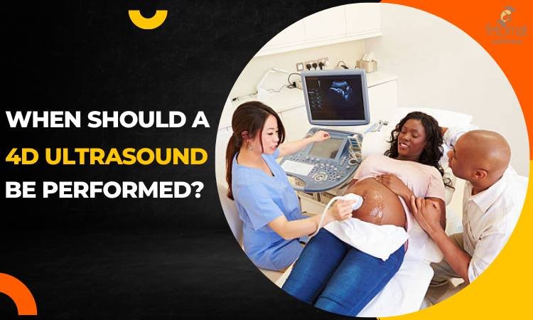
When is the Best Time to perform a 4D Ultrasound?
The ideal period to have a 4D ultrasound is between weeks 26 and 32, and the best times to watch in-depth ultrasound videos are weeks 27, 28 and 29. The best technique for diagnosing potential problems is the conventional 2D ultrasound.
The ideal thing to do is to:
- Perform a 4D echo during prenatal diagnosis and a 2D echo to fully assess the infant.
- Calculate weight to determine if the fetus is too “fat” or “thin”.
- Determine whether the placenta is functioning normally and whether there is enough amniotic fluid.
- Measure Doppler flows in the umbilical, cerebral, uterine, and other arteries.
A mother finds comfort in the 4D ultrasound since she can see her baby clearly and because a prenatal diagnosis specialist has thoroughly examined her kid. 4d ultrasound early pregnancy Kolkata is quite common due to this reason, but it should always be conducted at a reputed clinic where doctors and staffs are knowledgeable and properly equipped to conduct this type of scan.
Is there a risk associated with 4D ultrasound?
The question of whether ultrasounds can cause harm to a fetus is one that is frequently asked. The answer to this is that it is a straightforward, harmless approach that causes no pain. Because the baby does not experience an ultrasound’s theoretical heat, it is a safe procedure for both the expectant mother and the unborn child. Modern ultrasound probes are made such that the fetus will not be affected by microwaves.
Ultrasounds may be performed weekly or every three months. There is no need to perform more than a conventional 2D ultrasound during each trimester of pregnancy in 80% of cases, with the exception of couples who want to have a 4D echo within weeks 26 – 32. These situations must be handled privately because social security does not provide coverage.
Recommended Article About 4D Ultrasound: Ultrasounds During Pregnancy: All You Need to Know
How is 4D Ultrasound Conducted?
You can use an abdominal or vaginal ultrasound. Because it provides a sharper view of the abdominal route, vaginal 4D ultrasound is the preferred technique of most sonographers during the first few weeks of pregnancy and to measure the cervix (in cases where pregnancies are suspected of being at risk of premature labor or miscarriage). The approach that is most frequently employed is the abdominal one.